Posted by
Shazy on Saturday, February 26, 2011
Wellcome Image Awards 2011
 |
| Wellcome Image Awards 2011 |
Striking microscope images nab awardsColorful shots of a ruby-tailed wasp and a human chromosome bring mostly unseen objects to life.
 |
| Wellcome Image Awards 2011 |
Wellcome Images is world's leading source of ...
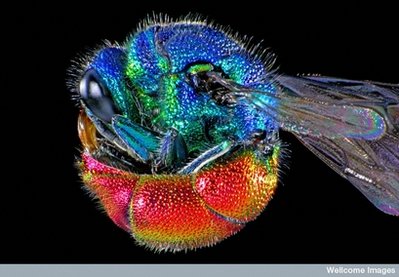
Wellcome Images is the world's leading source of images of medicine and its history. The Wellcome Image Awards recognize the creators of the most informative, striking and technically excellent images among recent acquisitions. You can view all of the 2011 winners at http://www.wellcomeimageawards.org/. This photomicrograph shows an adult ruby-tailed wasp curled into a ball. The wasp was lit with two electronic flashes while imaging to highlight the natural iridescent colours on its body. |
| Wellcome Image Awards 2011 |
Photomicrograph of a mosquito from the Culex ...
Photomicrograph of a mosquito from the Culex genus. This image shows the whole body of an adult male. The sample is from a microscope slide created in the middle of the 20th century. |
| Wellcome Image Awards 2011 |
Cell division and gene expression
Cell division and gene expression in plant cells. Confocal micrograph showing the expression of different fluorescent proteins in the stem of a thale cress seedling (Arabidopsis thaliana). Arabidopsis was the first plant to have its entire genome sequenced and is an important model for studying plant biology. The middle of the image shows a region of high cell proliferation, which drives the growth and branching of the seedling. |
| Wellcome Image Awards 2011 |
Polarised photomicrograph showing rows of suckers
Polarised photomicrograph showing the rows of suckers on the foreleg of a male diving beetle. Commonly known as the great diving beetle, these are largest freshwater beetles in the UK. They have a large streamlined body that is dark brown in colour, with a yellow abdomen and yellow legs. This image was produced by passing light through coloured filters, a technique known as Rheinberg illumination. |
| Wellcome Image Awards 2011 |
Cavefish embryo at around five days post-fertilisation. ...
Cavefish embryo at around five days post-fertilisation. The embryo has been stained with an antibody against a calcium-binding protein (in green) to show different neuronal types and their processes in the nervous system. This staining also reveals taste buds, which are located around the mouth and along the body of the cavefish. |
| Wellcome Image Awards 2011 |
Honey Bee - False colour scanning electron micrograph ...
Honey Bee - False colour scanning electron micrograph of a Honey bee. The honeybee has a hairy thorax and segented abdomen, a pair of double wings and three pairs of segmented legs, each one with a different 'tool', designed for a specific functions to assist in the collection and transport of pollen. |
| Wellcome Image Awards 2011 |
Image of human chromosome in metaphase
Image of a human chromosome in metaphase. The colours in this image indicate the density of chromatin in the chromosome, like a heat map (red shows high density, blue low). A low density of chromatin indicates a high level of gene expression, and a high density indicates repression of gene expression.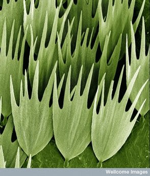 |
| Wellcome Image Awards 2011 |
Moth wing scales
Moth wing scales. Scanning electron micrograph (SEM) of the scales on the wing of a Madagascan moon moth, Argema mittrei. This moth is also known as the Comet moth, after its very long tail. The tail span is 15 cm and wing span 20 cm, making it one of the world's largest silk moths. |
| Wellcome Image Awards 2011 |
This colour-enhanced photomicrograph shows different ...
This colour-enhanced photomicrograph shows different species of bacteria that cause dental plaque - a colourless film that forms on teeth caused by the growth of bacterial colonies. The sample was removed from the mouth of a patient diagnosed with an aggressive form of gum disease.